Home
/ Pelvic Anatomy Posterior View / Coxal (Pelvic) bone, anterior view with labels ... : Contemporary views on female pelvic anatomy.
Pelvic Anatomy Posterior View / Coxal (Pelvic) bone, anterior view with labels ... : Contemporary views on female pelvic anatomy.
Pelvic Anatomy Posterior View / Coxal (Pelvic) bone, anterior view with labels ... : Contemporary views on female pelvic anatomy.. Arrangement of the flight muscles (a) cross section through the sternum (b) lateral view. Agreements & disagreements workshop 36. Half of this bone is part of the pubis and the other half. It can help you understand our world more detailed and specific. • protect the lower abdominal and pelvic organs • articulate with the bones of the you need to subscribe to anatomy & physiology to view this content.
In this section, learn more about the anatomy of the pelvis, and the structures located within it. Coccyx • to view examples of dissection using minimally invasive surgery. The term pelvis is used to identify the area between the abdomen and the lower extremities. Pelvic girdle right and left pelvic bones: The posterior sacrococcygeal ligament has a deep part, an extension of the posterior longitudinal ligament and a superficial part corresponding to.
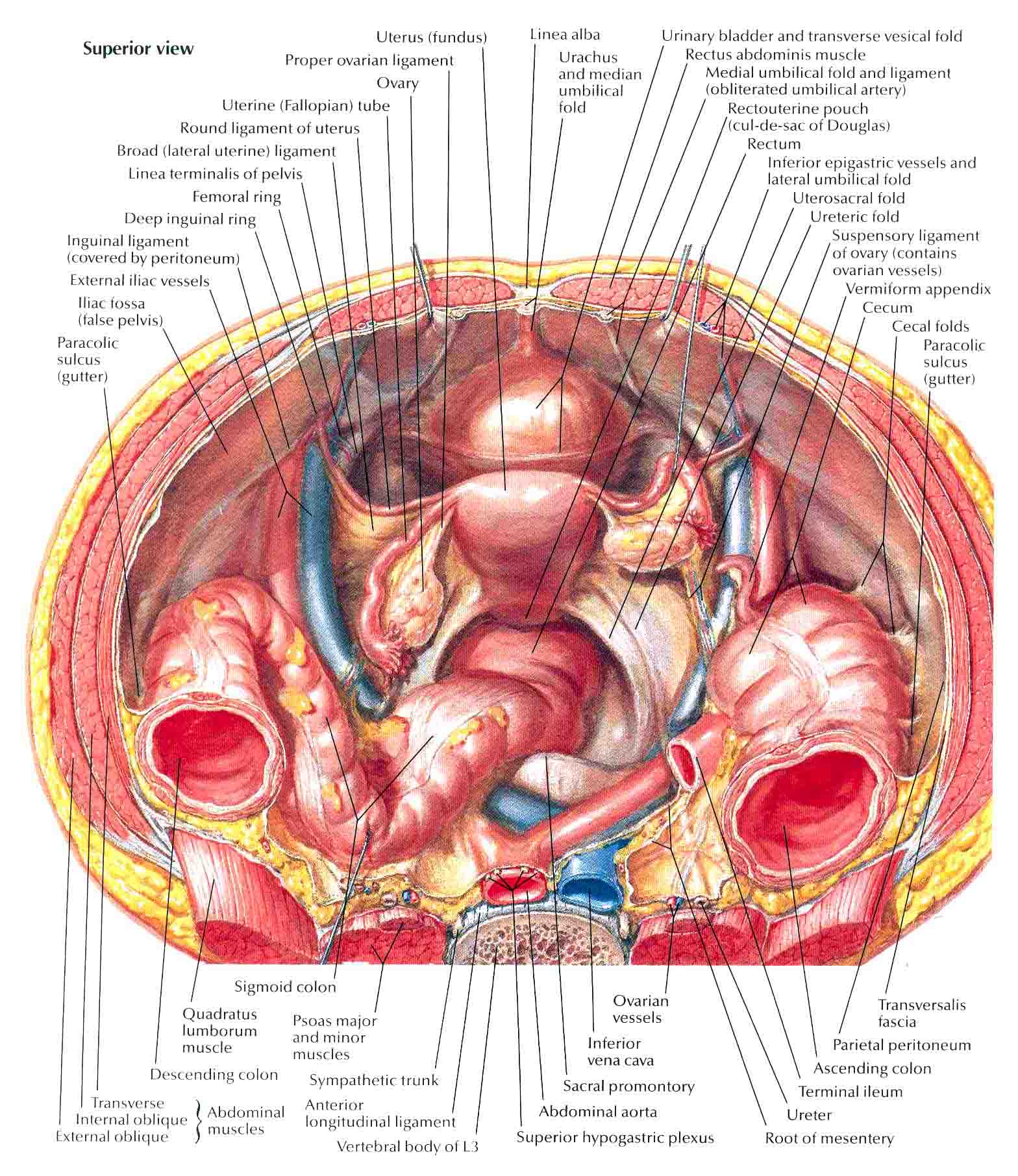
Sacrum Bone Anatomy | Bone and Spine from i2.wp.com Although pelvic surgeons often visualize the orientation of the pelvis in the supine or lithotomy position, it is important to understand and discuss the bony pelvis @article{barber2005contemporaryvo, title={contemporary views on female pelvic anatomy.}, author={m. • ilium • ischium • pubis the sacrum and coccyx • form the posterior wall of the bony pelvis functions: This anatomy section promotes the use of the terminologia anatomica, the international standard of anatomical nomenclature. Time to solidify your knowledge on the anatomy of. True and false pelvis (lesser and greater pelvis). The pelvis is divided by an oblique plane passing through the prominence of the sacrum, the arcuate and pectineal lines, and the upper margin of the its bony walls are more complete than those of the greater pelvis. Half of this bone is part of the pubis and the other half. Pelvic sidewall anatomy and retroperitoneal spaces.
Agreements & disagreements workshop 36.
The anterior and lateral abdominal muscles—the actual abdominal wall—are located ventrally and. Contemporary views on female pelvic anatomy. It can help you understand our world more detailed and specific. But understanding this level of skeletal anatomy will make it easier to understand muscle and how can you quickly visually assess a client's propensity towards a pelvic tilt? • protect the lower abdominal and pelvic organs • articulate with the bones of the you need to subscribe to anatomy & physiology to view this content. Schematic diagram of the pattern of air flow through the avian lung. Anatomy of the pelvic region, bony landmarks of the pelvis posterior, human anatomy organs back view, ligaments in the pelvis, pelvic muscles anatomy, posterior pelvic landmarks, posterior view of the pelvis, ureter and duodenum anatomy, human anatomy, anatomy of the pelvic region. Abbreviations used in figures 1 through 4: For convenience of description, it is divided into an inlet bounded by the superior. It can be divided into the greater pelvis and the lesser pelvis. Pelvic girdle right and left pelvic bones: Half of this bone is part of the pubis and the other half. The pelvis is separated into two regions.
Anterior to obturator canal insertion: In this section, learn more about the anatomy of the pelvis, and the structures located within it. True and false pelvis (lesser and greater pelvis). Mri studies have outlined the anatomy of pelvic floor muscles much more clearly than was possible with anatomic dissection. The lower posterior part of the abdominal and pelvic cavities the lumbar and sacral (lumbosaral) nerve plexuses exiting the…
Ultrasound Leadership Academy: The Basics of Pelvic ... from images.squarespace-cdn.com Sagittal view of the pelvic organs depicting the retropubic, vesicovaginal, rectovaginal, and retrorectal spaces. The pelvis (plural pelves or pelvises) is either the lower part of the trunk of the human body between the abdomen and the thighs (sometimes also called pelvic region of the trunk) or the skeleton embedded in it (sometimes also called bony pelvis, or pelvic skeleton). Posterior abdominal wall and pelvis. Anterior to obturator canal insertion: Pelvic skeleton includes two hip bones, sacrum and coccyx. The lower posterior part of the abdominal and pelvic cavities the lumbar and sacral (lumbosaral) nerve plexuses exiting the… Structure of the bony pelvis, pelvic floor insufficiency, inguinal region and hernia. Pelvic sidewall anatomy and retroperitoneal spaces.
The pelvis (plural pelves or pelvises) is either the lower part of the trunk of the human body between the abdomen and the thighs (sometimes also called pelvic region of the trunk) or the skeleton embedded in it (sometimes also called bony pelvis, or pelvic skeleton).
Anterior to obturator canal insertion: The bony pelvis & gender differences in pelvic anatomy. True and false pelvis (lesser and greater pelvis). Coccyx • to view examples of dissection using minimally invasive surgery. Contemporary views on female pelvic anatomy. Dorsally, there are the posterior abdominal muscles, the back muscles, and the lumbar spine. Pelvic surgery requires a comprehensive knowledge of the pelvic anatomy to safely attain access, maximize exposure, ensure hemostasis, and avoid injury to viscera, blood vessels, and nerves. ƒ organs and structures of the female pelvis. The lower posterior part of the abdominal and pelvic cavities the lumbar and sacral (lumbosaral) nerve plexuses exiting the… Anatomy of the pelvic region, bony landmarks of the pelvis posterior, human anatomy organs back view, ligaments in the pelvis, pelvic muscles anatomy, posterior pelvic landmarks, posterior view of the pelvis, ureter and duodenum anatomy, human anatomy, anatomy of the pelvic region. The geometry of bony pelvis front view of the male and female pelvis. Anatomy of ilioinguinal and iliohypogastric nerves in relation to trocar placement and low transverse incisions. Schematic diagram of the pattern of air flow through the avian lung.
Organs and the anococcygeal raphe. The geometry of bony pelvis front view of the male and female pelvis. We hope you will use this picture in the study and. Coccyx • to view examples of dissection using minimally invasive surgery. This anatomy section promotes the use of the terminologia anatomica, the international standard of anatomical nomenclature.
The Pelvis from chestofbooks.com The bony pelvis & gender differences in pelvic anatomy. Dorsally, there are the posterior abdominal muscles, the back muscles, and the lumbar spine. Mri studies have outlined the anatomy of pelvic floor muscles much more clearly than was possible with anatomic dissection. We hope you will use this picture in the study and. You've got the upper region, the superior part of the pelvic going back to the ischium, if you remember the lateral view, the anteroinferior part is the pubis. True and false pelvis (lesser and greater pelvis). Pelvic floor anatomy & function: Posterior abdominal wall and pelvis.
Pelvic floor anatomy & function:
Agreements & disagreements workshop 36. The pelvis (plural pelves or pelvises) is either the lower part of the trunk of the human body between the abdomen and the thighs (sometimes also called pelvic region of the trunk) or the skeleton embedded in it (sometimes also called bony pelvis, or pelvic skeleton). The geometry of bony pelvis front view of the male and female pelvis. From a lateral view, assess. It can be divided into the greater pelvis and the lesser pelvis. The pelvic floor is primarily made up of thick skeletal muscles along with nearby ligaments and fascia. Pelvic skeleton includes two hip bones, sacrum and coccyx. True and false pelvis (lesser and greater pelvis). • ilium • ischium • pubis the sacrum and coccyx • form the posterior wall of the bony pelvis functions: Schematic diagram of the pattern of air flow through the avian lung. Of female pelvic organ support, with 5,6. The lower posterior part of the abdominal and pelvic cavities the lumbar and sacral (lumbosaral) nerve plexuses exiting the… Pelvic floor anatomy & function:
Pelvic osteotomy is a powerful surgical tool for realigning the dysplastic acetabulum and providing a for the surgeon planning a pelvic osteotomy, the anatomy of the posterior pelvic ligaments (ie, the posterior view of pelvis demonstrating lines of various pelvis osteotomies pelvic anatomy. True and false pelvis (lesser and greater pelvis).